Why BDAT matters?
Portable: it can be used at the patients in hospital, in local health centers or even at home.
Fast: each measurement takes about 5 minutes, thanks to a robust protocol and guiding interface.
Inclusive: brings advanced bone diagnostics to regions where DXA is not widely available.

Clinical use of BDAT at the distal forearm (radius) — a quick and non-invasive exam (source: Reasearch features - 2023).
Beyond research labs, BDAT is part of a wider movement to make health technology accessible and preventive. In Chile, for example, local hospitals such as Hospital Gustavo Fricke have joined the project to screen older adults at risk of fracture. According to the national press (El Mostrador, 2023), this collaboration aims to develop a low-cost, portable solution that could help thousands of Chileans —especially those living far from major hospitals— to monitor their bone quality early and avoid future fractures.
The International Bone Ultrasound Society (BoneUs) promotes the development and progress of bone QUS in its different aspects, its technologies and applications.
The long-term vision is clear: a world where anyone, anywhere, can have their bone quality checked with a simple, safe, and affordable ultrasound test.
Sources:
Research Features (2023) - “Jean-Gabriel Minonzio: Ultrasonic assessment of bone fragility” (English).
El Mostrador (2023) - “El innovador estudio chileno de densitometría ósea a través de ultrasonido” (Spanish).
Universidad de Valparaiso (2024) - "Con ultrasonido y biopsia, académico de Informática UV busca diferenciar fracturas por fragilidad de cadera" (Spanish).
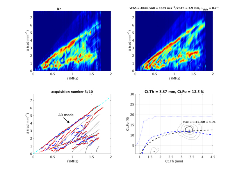
A dedicated ultrasonic probe is positioned at the distal one-third of the radius with coupling gel. The system acquires multiple bidirectional transmissions to extract first-arrival and guided-wave mode velocities (vFAS, vA0) and builds a guided-wave spectral image for inversion, providing cortical thickness (Ct.Th) and cortical porosity (Ct.Pobcm). A standard acquisition protocol collects ~10 captures and retains coherent series for analysis. The JoVE article provides step-by-step guidance for operator positioning and quality checks.
Dubot, B., Deniau, M., Ramiandrisoa, D., Minonzio, J. Cortical Bone Assessment Using Ultrasonic Guided Waves: A Reproducibility Study in a Healthy Population. J. Vis. Exp. (215), e66985,(2025). DOI: 10.3791/66985.
Dubot, B., Deniau, M., Ramiandrisoa, D., Minonzio, J. Cortical Bone Assessment Using Ultrasonic Guided Waves: A Reproducibility Study in a Healthy Population. J. Vis. Exp. (215), e66985,(2025). DOI: 10.3791/66985.
Clinical Studies & Validation

References:
[1] Talmant M., Kolta S., Roux C., Haguenauer D., Bossy E., Laugier P. In vivo performance evaluation of bi-directional ultrasonic axial transmission for cortical bone assessment. Ultrasound in Medicine & Biology, 2009. DOI: 10.1016/j.ultrasmedbio.2008.12.008
[2] Schneider J., Raum K., Pumberger M., Zippelius T., Hoff E., Strube P., Putzier M., Minonzio J.-G., Laugier P. Prospective discrimination of vertebral fractures by axial transmission ultrasound using optimized first arriving signal velocity measurements. IEEE International Ultrasonics Symposium (IUS), 2015. DOI: 10.1109/ULTSYM.2015.0513
[3] Schneider J., Varga P., Raum K., Pumberger M., Zippelius T., Hoff E., Strube P., Putzier M., Minonzio J.-G., Laugier P. Multisite ultrasound axial transmission study in postmenopausal women using optimized first arriving signal velocity measurements. European Symposium on Ultrasonic Characterization of Bone (ESUCB), 2015. DOI: 10.1109/ESUCB.2015.7169895
[4] Minonzio J.-G., Bochud N., Vallet Q., Chappard C., Talmant M., Laugier P. Ultrasound-based estimates of cortical bone thickness and porosity are associated with nontraumatic fractures in postmenopausal women: a pilot study. Journal of Bone and Mineral Research, 2019. DOI: 10.1002/jbmr.3733
[5] Minonzio J.-G., Ramiandrisoa D., Schneider J., Kohut E., Streichhahn M., Stervbo U., et al. Bi-Directional Axial Transmission measurements applied in a clinical environment. PLoS ONE, 2022. DOI: 10.1371/journal.pone.0277831 — Data available at: 10.17605/OSF.IO/XY9QV
[6] Rojo F., Schneider J., Minonzio J.-G., Laugier P., Vallet Q. Classification of hip fragility fractures in older adults using an ultrasonic device. IEEE International Ultrasonics Symposium (IUS), 2023. DOI: 10.1109/IUS51837.2023.10307317

References:
[1] Mitton D., Minonzio J.-G., Talmant M., Ellouz R., Rongieras F., Laugier P., Bruyère-Garnier K. Non-destructive assessment of human ribs mechanical properties using quantitative ultrasound. Journal of Biomechanics, 2014. DOI: 10.1016/j.jbiomech.2014.01.052
[2] Minonzio J.-G., Zapata E., Bochud N., Vallet Q., Rongieras F., Pialat J.-B. Ex vivo radius fracture discrimination from cortical thickness and porosity obtained by axial transmission. IEEE International Ultrasonics Symposium (IUS), 2018. DOI: 10.1109/ULTSYM.2018.8579720
[3] Minonzio J.-G., Bochud N., Vallet Q., Bala Y., Ramiandrisoa D., Follet H., Mitton D., Laugier P. Cortical thickness and porosity assessment using ultrasound guided waves: an ex vivo validation. Bone, 2018. DOI: 10.1016/j.bone.2018.07.018
[4] Schneider J., Iori G., Ramiandrisoa D., Hammami M., Gräsel M., Chappard C., Barkmann R., Laugier P., Grimal Q., Minonzio J.-G., Raum K. Ex vivo cortical porosity and thickness predictions at the tibia using full-spectrum ultrasonic guided-wave analysis. Archives of Osteoporosis, 2019. DOI: 10.1007/s11657-019-0578-1

References:
[1] Vallet Q., Bochud N., Chappard C., Laugier P., Minonzio J.-G. In vivo characterization of cortical bone using guided waves measured by axial transmission. IEEE Transactions on Ultrasonics, Ferroelectrics, and Frequency Control, 2016. DOI: 10.1109/TUFFC.2016.2587079
[2] Schneider J., Ramiandrisoa D., Armbrecht G., Ritter J., Felsenberg D., Raum K., Minonzio J.-G. In vivo measurements of cortical thickness and porosity at the proximal third of the tibia using guided waves: comparison with site-matched pQCT and distal HR-pQCT. Ultrasound in Medicine & Biology, 2019. DOI: 10.1016/j.ultrasmedbio.2019.01.008
[3] Behforootan S., Thorniley M., Minonzio J.-G., Boughton O., Karia M., Bhattacharya R., Hansen U., Cobb J., Abel R. Can guided wave ultrasound predict bone mechanical properties at the femoral neck in patients undergoing hip arthroplasty. Journal of the Mechanical Behavior of Biomedical Materials, 2022. DOI: 10.1016/j.jmbbm.2022.105468
[4] Dubot B., Deniau M., Ramiandrisoa D., Minonzio J.-G. Cortical Bone Assessment Using Ultrasonic Guided Waves: A Reproducibility Study in a Healthy Population. Journal of Visualized Experiments, 2025. DOI: 10.3791/66985
[5] Minonzio J.-G., Ramiandrisoa D., Fernandez S., Chappard C., Cohen-Solal M. Cortical bone parameters measured at the one-third distal radius using BDAT and HR-pQCT. Ultrasonics, 2025. DOI: 10.1016/j.ultras.2025.107829 — Data available at : https://osf.io/ehqrb
Methods

References:
[1] Minonzio J.-G., Talmant M., Laugier P. Guided wave phase velocity measurement using multi-emitter and multi-receiver arrays in the axial transmission configuration. Journal of the Acoustical Society of America, 2010. DOI: 10.1121/1.3377085
[2] Xu K., Ta D., Cassereau D., Hu B., Wang W., Laugier P., Minonzio J.-G. Multichannel processing for dispersion curves extraction of ultrasonic axial-transmission signals. Journal of the Acoustical Society of America, 2016. DOI: 10.1121/1.4962491
[3] Xu K., Minonzio J.-G., Ta D., Hu B., Wang W., Laugier P. Sparse inverse SVD method for high-resolution extraction of the dispersion curves of ultrasonic guided waves. IEEE Transactions on Ultrasonics, Ferroelectrics, and Frequency Control, 2016. DOI: 10.1109/TUFFC.2016.2592688
[4] Xu K., Laugier P., Minonzio J.-G. Dispersive Radon transform. Journal of the Acoustical Society of America, 2018. DOI: 10.1121/1.5036726
[5] Bai L., Xu K., Bochud N., Ta D., Hu B., Laugier P., Minonzio J.-G. Multichannel wideband mode-selective excitation of ultrasonic guided waves in long cortical bone. Proceedings of the IEEE International Ultrasonics Symposium (IUS), 2016. DOI: 10.1109/ULTSYM.2016.7728774
[6] Bossy E., Talmant M., Defontaine M., Patat F., Laugier P. Bidirectional axial transmission can improve accuracy and precision of ultrasonic velocity measurement in cortical bone: a validation on test materials. IEEE Transactions on Ultrasonics, Ferroelectrics, and Frequency Control, 2004. DOI: 10.1109/TUFFC.2004.1268469
[7] Moreau L., Minonzio J.-G., Foiret J., Bossy E., Talmant M., Laugier P. Accurate measurement of guided modes in a plate using a bidirectional approach. Journal of the Acoustical Society of America, 2014. DOI: 10.1121/1.4832335
[8] Chen J., Foiret J., Minonzio J.-G., Talmant M., Su Z., Cheng L., Laugier P. Measurement of guided mode wavenumbers in soft tissue–bone mimicking phantoms using ultrasonic axial transmission. Physics in Medicine and Biology, 2012. DOI: 10.1088/0031-9155/57/10/3025
[9] Bochud N., Vallet Q., Minonzio J.-G., Laugier P. Predicting bone strength with ultrasonic guided waves. Scientific Reports, 2017. DOI: 10.1038/srep43628
[10] Minonzio J.-G., Foiret J., Talmant M., Laugier P. Impact of attenuation on guided mode wavenumber measurement in axial transmission on bone-mimicking plates. Journal of the Acoustical Society of America, 2011. DOI: 10.1121/1.3652884
[11] Moreau L., Minonzio J.-G., Talmant M., Laugier P. Measuring the wavenumber of guided modes in waveguides with linearly varying thickness. Journal of the Acoustical Society of America, 2014. DOI: 10.1121/1.4869691
[12] Minonzio J.-G., Foiret J., Moilanen P., Pirhonen J., Zhao Z., Talmant M., Timonen J., Laugier P. A free plate model can predict guided modes propagating in tubular bone-mimicking phantoms. Journal of the Acoustical Society of America, 2015. DOI: 10.1121/1.4903920

References:
[1] Talmant M., Foiret J., Minonzio J.-G. Guided Waves in Cortical Bone. Bone Quantitative Ultrasound (book chapter), 2011. DOI: 10.1007/978-94-007-0017-8_7
[2] Granke M., Grimal Q., Saïed A., Nauleau P., Peyrin F., Laugier P. Change in porosity is the major determinant of the variation of cortical bone elasticity at the millimeter scale in aged women. Bone, 2011. DOI: 10.1016/j.bone.2011.08.002
[3] Foiret J., Minonzio J.-G., Chappard C., Talmant M., Laugier P. Combined estimation of thickness and velocities using ultrasound guided waves: a pioneering study on in vitro cortical bone samples. IEEE Transactions on Ultrasonics, Ferroelectrics, and Frequency Control, 2014. DOI: 10.1109/TUFFC.2014.3062
[4] Bochud N., Vallet Q., Bala Y., Follet H., Minonzio J.-G., Laugier P. Genetic algorithms-based inversion of multimode guided waves for cortical bone characterization. Physics in Medicine and Biology, 2016. DOI: 10.1088/0031-9155/61/19/6953
[5] Bochud N., Vallet Q., Minonzio J.-G., Laugier P. Predicting bone strength with ultrasonic guided waves. Ultrasound Med. Biol. 2017. DOI: 10.1038/srep43628
[6] Minonzio J.-G., Bochud N., Vallet Q., Bala Y., Ramiandrisoa D., Follet H., Mitton D., Laugier P. Bone cortical thickness and porosity assessment using ultrasound guided waves: an ex vivo validation study. Bone, 2018. DOI: 10.1016/j.bone.2018.07.018
[7] Araya C., Martinez A., Ramiandrisoa D., Ta D., Xu K., Osses A., Minonzio J.-G. Real-time waveguide parameter estimation using axial transmission SVD-based method and sparse dispersive Radon transform. Proceedings of the IEEE Latin America Ultrasonics Symposium (LAUS), 2021. DOI: 10.1109/LAUS53676.2021.9639171
[8] Aróstica R., Aguilera A., Osses A., Minonzio J.-G. A simplified homogenization model applied to viscoelastic cortical bone at ultrasonic frequencies. Journal of Biomechanics, 2022. DOI: 10.1016/j.jbiomech.2021.110868

References:
[1] Minonzio J.-G., Cataldo B., Olivares R., Ramiandrisoa D., Soto R., Crawford B., de Albuquerque V. H. C., Muñoz R. Automatic Classifying of Patients With Non-Traumatic Fractures Based on Ultrasonic Guided Wave Spectrum Image Using a Dynamic Support Vector Machine. IEEE Access, 2020. DOI: 10.1109/ACCESS.2020.3033480
[2] Miranda D., Olivares R., Muñoz R., Minonzio J.-G. Improvement of Patient Classification Using Feature Selection Applied to Bidirectional Axial Transmission. IEEE Transactions on Ultrasonics, Ferroelectrics, and Frequency Control, 2022. DOI: 10.1109/TUFFC.2022.3195477
[3] Flores Cisternas W., Aguilera A. I., Olivares R., Muñoz R., Minonzio J.-G. CNN applied to ultrasonic guided wave spectrum image classification. Journal of Physics: Conference Series, 2024. DOI: 10.1088/1742-6596/2822/1/012021
[4] Díaz D., Flores W., Aguilera A. I., Olivares R., Muñoz R., Minonzio J.-G. Fragility Fracture Classification Using Axial Transmission Raw Signals and Multi-Channel Convolutional Neural Network. Proceedings of the IEEE Latin America Ultrasonics Symposium (LAUS), 2024. DOI: 10.1109/LAUS60931.2024.10553065
Point transducer
Reference: Kassou K., Remram Y., Laugier P., Minonzio J.-G. Dispersion characteristics of the flexural wave assessed using low-frequency (50 to 150 kHz) point-contact transducers: A feasibility study on bone-mimicking phantoms.
Ultrasonics, 2017. DOI: 10.1016/j.ultras.2017.05.008
CMUT (capacitive micromachined ultrasonic transducer)
Reference: Boulmé A., Ngo S., Minonzio J.-G., Legros M., Talmant M., Laugier P., Certon D. A capacitive micromachined ultrasonic transducer probe for assessment of cortical bone. IEEE Transactions on Ultrasonics, Ferroelectrics, and Frequency Control, 2014. DOI: 10.1109/TUFFC.2014.2959
Pulse echo at 1/3 distal radius
Reference : Minonzio J.-G., Han C., Cassereau D., Grimal Q. In vivo pulse-echo measurement of apparent broadband attenuation and Q factor in cortical bone: a preliminary study. Physics in Medicine and Biology, 2021. DOI: 10.1088/1361-6560/ac1022
Minonzio J.-G., Talmant M., Laugier P., « Procédé et dispositif ultrasonores pour caractériser un milieu », FR2946753 (A1) / WO2010142927 (A1). WO2010142927A1.
Moreau L., Minonzio J.-G., Talmant M., Laugier P., « Procédé et dispositif ultrasonores pour représenter la propagation d'ondes ultrasonores dans un guide d'épaisseur linéairement variable », FR1357204 / WO2015010878 (A1). WO2015010878A1.
Minonzio J.-G., Xu K., Ta D., Bai L., « Method and device for characterizing a waveguide », EP17306027 / WO2019025510 (A1). WO2019025510A1
Ex vivo — Bone 2018
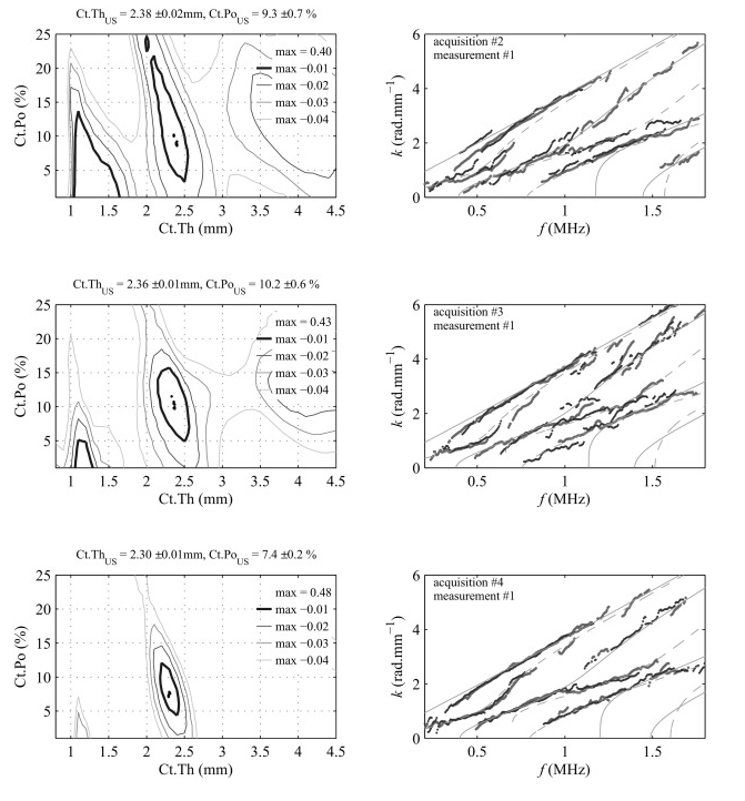
Left column: illustration of a typical objective function (Eq. (4)) for three acquisitions with intermediate repositioning on the radius specimen represented in Fig.
Right column: comparison between experimental dispersion curves (dots) and the optimal predicted modes (continuous and dashed lines). Values correspond to the mean and standard deviation of the ten measurements (black dots).
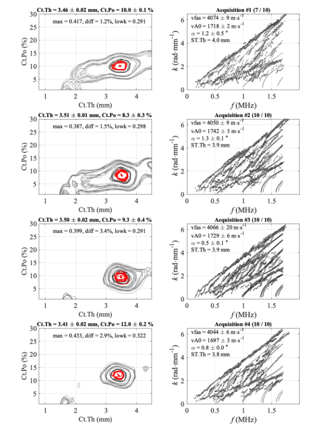
In vivo — JoVE 2025
The figure shows 4 series for 1 participant and 1 operator: inverse problem images (left column) and experimental wavenumbers vs best fitting model (right column).
Values correspond to the mean and standard deviation of the kept acquisitions over each 10-acquisition series. The number of kept acquisitions is shown in the title (e.g., 7/10).
References:
[1] Minonzio J.-G., Bochud N., Vallet Q., Bala Y., Ramiandrisoa D., Follet H., Mitton D., Laugier P. Cortical thickness and porosity assessment using ultrasound guided waves: an ex vivo validation. Bone, 2018. DOI: 10.1016/j.bone.2018.07.018
[2] Dubot B., Deniau M., Ramiandrisoa D., Minonzio J.-G. Cortical Bone Assessment Using Ultrasonic Guided Waves: A Reproducibility Study in a Healthy Population. Journal of Visualized Experiments, 2025. DOI: 10.3791/66985